Treatment only works when the diagnosis is correct. That sounds obvious. But you would be surprised how many couples spend months on the wrong treatment plan because the initial evaluation was incomplete or the scans were done carelessly. A poorly timed ultrasound. A missed hormonal marker. A follicle count done on the wrong cycle day. Small oversights that lead to big delays.
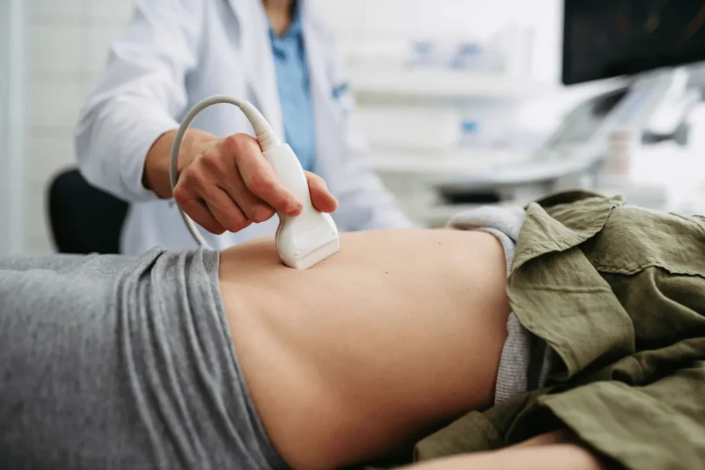
If you are looking for accurate sonography and fertility diagnostics in Wakad, Dr. Pavan Bendale (M.B.B.S., DGO, DNB) runs diagnostics the way they should be run. Thorough. Precisely timed. Interpreted by the same doctor who will be treating you. Not by a technician who hands you a printout and sends you on your way.
What Diagnostic Services Are Available?
Diagnostics at Dr. Pavan Bendale’s practice cover both fertility evaluation and general gynecological assessment. Here is the full range of what is offered:
1. Transvaginal Ultrasound (TVS)
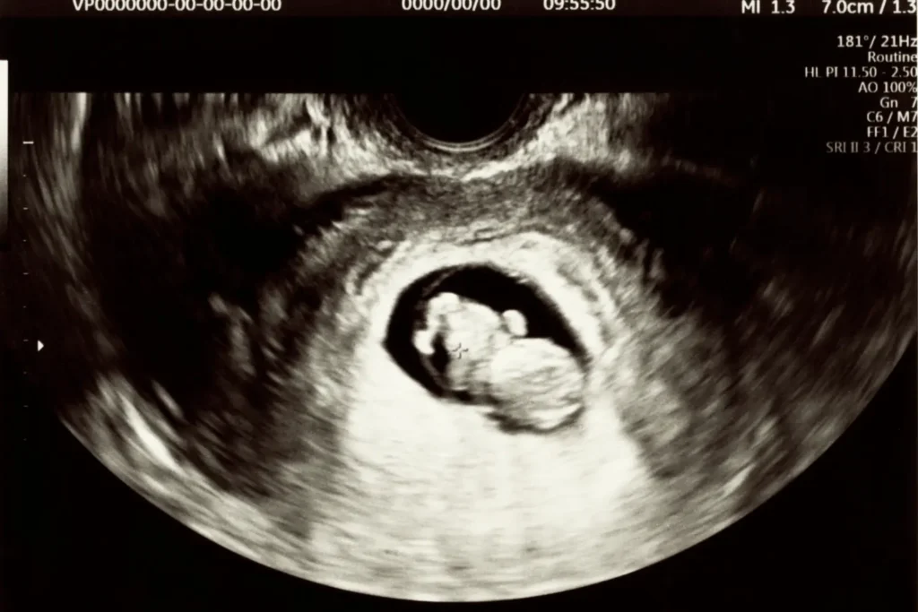
The most fundamental imaging tool in gynecology and fertility. A transvaginal probe provides high-resolution images of the uterus, ovaries, endometrial lining, and surrounding structures. It is used to detect ovarian cysts, fibroids, polyps, adenomyosis, congenital uterine anomalies, and to measure ovarian reserve through antral follicle count (AFC). Dr. Pavan Bendale performs the scan himself, which means the person interpreting the image is the same person making your treatment decisions.
2. Follicle Monitoring (Follicular Study)
This is the backbone of any medicated fertility cycle, whether you are doing timed intercourse, IUI, or IVF. Serial ultrasound scans track how your follicles are growing in response to ovulation induction medications. Each scan measures follicle size, number, and endometrial thickness. The data guides medication adjustments, trigger shot timing, and the optimal window for insemination or egg retrieval. Getting this wrong by even a day can cost you the entire cycle.
3. 3D and 4D Ultrasound
Three-dimensional ultrasound creates a detailed volumetric image of the uterus and ovaries. It is particularly valuable for diagnosing uterine anomalies like septate uterus, bicornuate uterus, or arcuate uterus that standard 2D scans can miss. 4D adds real-time movement, which is useful during pregnancy for assessing fetal anatomy and movement patterns. These scans provide diagnostic clarity that flat 2D images simply cannot match in complex cases.
4. Saline Infusion Sonography (SIS) / Sonohysterography
A small amount of sterile saline is infused into the uterine cavity during an ultrasound. This distends the cavity and creates contrast, making it far easier to identify intrauterine abnormalities like polyps, submucosal fibroids, adhesions (synechiae), or an irregular endometrial lining. SIS is less invasive than hysteroscopy and provides excellent diagnostic accuracy for cavity-related issues.
5. Doppler Ultrasound
Doppler scans assess blood flow to the uterus, ovaries, and during pregnancy, to the placenta and baby. In fertility, uterine artery Doppler helps evaluate endometrial receptivity before embryo transfer. During pregnancy, it monitors blood supply to the baby and detects early signs of placental insufficiency or intrauterine growth restriction (IUGR).
6. Hormonal Assessments
Imaging alone does not tell the complete story. Dr. Pavan Bendale pairs ultrasound findings with targeted blood work to create a comprehensive diagnostic picture:
a) AMH (Anti-Mullerian Hormone) to assess ovarian reserve
b) FSH and LH (Day 2/3) to evaluate ovarian function and detect conditions like PCOS
c) Estradiol to correlate with follicle development during monitored cycles
d) Progesterone (Day 21) to confirm whether ovulation has occurred
e) Thyroid panel (TSH, T3, T4) because even mild thyroid imbalance can affect fertility and pregnancy outcomes
f) Prolactin levels, especially when irregular periods or absent ovulation are present
g) Insulin and glucose testing for PCOS patients to assess metabolic involvement
According to the American College of Obstetricians and Gynecologists (ACOG), the combination of transvaginal ultrasound with targeted hormonal evaluation forms the gold standard for initial fertility assessment and ongoing cycle monitoring.
“I went to three different scan centres before coming to Dr. Pavan Bendale. Each report said something different about my ovarian cyst. He did the scan himself, showed me exactly what it was on the screen, and explained why the previous reports were inconsistent. That was the first time I actually understood my own diagnosis.”
— Verified patient, Google Reviews
Why Does It Matter Who Does Your Scan?
At most diagnostic centres, a sonography technician performs the scan and a radiologist writes the report. Neither of them knows your clinical history, your symptoms, or what specific question needs answering. The result is a generic report that your treating doctor then has to reinterpret.
At Dr. Pavan Bendale’s practice, the equation is different. He performs the scans himself. That means:
1. The person holding the probe already knows your medical history, your symptoms, and what he is specifically looking for
2. Findings are interpreted in real time, not days later on a printed report
3. Treatment decisions can be made immediately based on what the scan reveals
4. Subtle findings that a technician might overlook or underreport are caught because the clinical context is already in mind
5. You get your answers in the same appointment, not after a chain of referrals and follow-ups
This is especially critical during follicle monitoring cycles where timing decisions need to happen within hours, not days. A delayed or misread scan can mean a missed ovulation window.
Dr. Pavan Bendale vs. Typical Diagnostic Centres: Comparison
| What Matters | Dr. Pavan Bendale | Typical Scan Centres |
|---|---|---|
| Doctor performs the scan personally | ✓ Dr. Pavan Bendale himself | ✗ Technician performs, radiologist reports |
| Scan interpreted with clinical context | ✓ Full patient history known | ✗ Generic reporting |
| Real-time treatment decisions | ✓ Same appointment | ✗ Report sent to referring doctor later |
| 3D/4D ultrasound available | ✓ On-site | ✗ Not always available |
| Saline infusion sonography (SIS) | ✓ Performed in-house | ✗ Rarely offered |
| Doppler assessment for fertility | ✓ Uterine and ovarian Doppler | ✗ Only basic pregnancy Doppler |
| Hormonal blood work paired with imaging | ✓ Integrated assessment | ✗ Scan only, blood work done separately |
| Follicle monitoring precision | ✓ Every 2-3 days, medication adjusted same day | ✗ Scans scheduled rigidly, delays in reporting |
Sonography During Pregnancy: What Scans Do You Need and When?
Beyond fertility, Dr. Pavan Bendale provides a complete pregnancy ultrasound schedule. Each scan has a specific purpose at a specific gestational window:
1. Early Pregnancy Scan (6 to 8 weeks)
Confirms intrauterine pregnancy, detects heartbeat, rules out ectopic pregnancy, and establishes gestational age. Particularly important for IVF pregnancies.
2. NT Scan with Dual Marker (11 to 13 weeks)
Nuchal translucency measurement combined with blood markers screens for chromosomal abnormalities like Down syndrome. This is a time-sensitive scan that must happen within this specific window.
3. Anomaly Scan / TIFFA (18 to 22 weeks)
The most detailed scan of the pregnancy. Every major organ system of the baby is examined. Brain, spine, heart, kidneys, limbs, face, and placental position are all assessed. This scan is non-negotiable.
4. Growth Scan (28 to 32 weeks)
Checks fetal growth, amniotic fluid levels, placental health, and baby’s position. For high-risk pregnancies, growth scans may be repeated more frequently with Doppler assessment.
5. Pre-Delivery Scan (36 to 38 weeks)
Final assessment of baby’s weight, position, cord status, and amniotic fluid before delivery planning. Helps determine whether vaginal delivery or planned caesarean is the safer option.
The WHO Recommendations on Antenatal Care support a minimum of one ultrasound before 24 weeks for all pregnancies, with additional scans as clinically indicated for higher-risk cases.
Frequently Asked Questions About Sonography and Diagnostics in Wakad
Is transvaginal ultrasound painful?
It causes mild discomfort, similar to a pelvic exam. The probe is slim and the procedure takes only a few minutes. Most patients find it far less uncomfortable than they expected.
How many follicle monitoring scans will I need per cycle?
Typically 3 to 4 scans per medicated cycle, spaced 2 to 3 days apart. The exact number depends on how your follicles respond to medication. Dr. Pavan Bendale adjusts the schedule based on real-time findings.
Can ultrasound detect all uterine problems?
Standard 2D ultrasound catches most issues. But some conditions like a uterine septum or small polyps are better identified with 3D ultrasound or saline infusion sonography. Dr. Pavan Bendale uses the appropriate imaging modality based on what your case requires.
Do I need to prepare for a sonography appointment?
For a transvaginal scan, no special preparation is needed. For some abdominal scans, you may be asked to come with a full bladder. The team provides clear instructions when you book your appointment.
Why should my treating doctor do the scan instead of a separate centre?
Because context matters. A technician sees an image. Your treating doctor sees an image connected to your symptoms, your history, your treatment plan, and your goals. That connection is what turns a scan from a picture into a clinical decision.
Are pregnancy scans safe for the baby?
Yes. Ultrasound uses sound waves, not radiation. It has been used in obstetrics for over 50 years with an excellent safety record. There is no evidence of harm to the mother or baby from diagnostic ultrasound when used appropriately.
“During my IUI cycle, I was getting follicle scans at an outside centre. The reports kept saying one follicle at 14mm. Dr. Pavan Bendale did the scan himself and found two follicles, one at 16mm and another hiding behind the ovary. That second follicle changed the entire treatment plan. Details matter, and he catches them.”
— Verified patient, Google Reviews
Accurate Diagnosis Is Not a Luxury. It Is the Starting Point.
Whether you are beginning your fertility evaluation, in the middle of a treatment cycle, or monitoring a pregnancy, the quality of your diagnostics determines the quality of every decision that follows.
Dr. Pavan Bendale performs and interprets your scans with the clinical context that only your treating doctor can bring. Paired with targeted hormonal assessments, this gives you the most complete and accurate picture of where you stand and what needs to happen next.
Dr. Pavan Bendale
301, 3rd Floor, Darekar Heights, Dange Chowk Road, Bhumkar Chowk Rd, Opp. Pandit Petrol Pump, Tathawade, Pune 411033
Call / WhatsApp: 07840950737
Email: pavanbendale007@gmail.com
See clearly. Decide confidently. Book your scan today.